| 6:00 - 13:25
|
|
Virtual Help Desk
|
| 7:00 - 7:15
|
|
Welcome and Opening Remarks
Session Chairs:
Andrew Yurochko, IHW 2021 Co-Chair
LSU Health Shreveport, United States
|
Blossom Damania, IHW 2021 Co-Chair
UNC - Chapel Hill, United States
|
Ian Mohr, IHW 2021 Co-Chair
NYU Grossman School of Medicine, United States
|
|
| 7:15 - 7:55
|
|
Plenary 1 - Viral Entry and Replication
Session Chairs:
Benedikt Kaufer, Project Director
Freie Universität Berlin, Germany
|
Britt Glaunsinger, Professor
University of California Berkeley Research, United States
|
Presenter:

|
Matthew Weitzman, Professor
University of Pennsylvania and Children’s Hospital of Philadelphia, United States
Biography
Matthew Weitzman grew up in Britain, graduating with a degree in Genetics from Leeds University and a PhD in Molecular Virology from Oxford Brookes. He was a Fogarty Fellow at the U.S. National Institutes of Health and was also a Postdoctoral Fellow at the University of Pennsylvania. He started his independent lab as a faculty member of the Salk Institute in La Jolla, California and University of California, San Diego. In 2011 he moved to his current position where he is Professor at the University of Pennsylvania and runs a lab at the Children’s Hospital of Philadelphia. His lab employs techniques in biochemistry, genetics and cell biology to harness human DNA viruses as model systems to investigate fundamental cellular processes.
|
HSV-1 Manipulation of the Host Cell Proteome
Herpesvirus infections are associated with extensive remodeling of the cellular proteome and nuclear architecture. During productive lytic infection these viruses harness cellular processes, while also counteracting host defenses. This is achieved by altering abundance, post-translational modifications, and localization of cellular proteins. Viral DNA replication takes place at nuclear assemblies termed viral replication compartments (VRCs). Cellular proteins exploited for viral replication are recruited to these compartments, while host antiviral proteins that antagonize viral infection are prevented from associating with viral genomes. Virus infection also activates cellular signaling pathways that promote replication, such as the DNA damage response. Viral gene products can manipulate host proteins to redirect cellular processes or subvert antiviral responses. We have developed integrated proteomics approaches to quantitate protein abundance, to detect changes in post-translational modifications such as phosphorylation and ubiquitination, and to identify protein associations with viral DNA during infection. Using ICP0 as an example early protein that alters the host proteome to promote infection and counter intrinsic host defenses, we integrated proteomics datasets to identify cellular substrates degraded in an ICP0-dependent manner or associated with the viral genome in the absence of ICP0. Our comparative proteomics analysis reveals cellular processes that are manipulated during HSV-1 infection and points to host factors counteracted by ICP0 protein as the host proteome is remodeled to promote efficient infection.
|
|
| 7:55 - 8:20
|
|
Symposium 1A
Presenter:

|
Thomas Stamminger, Prof. Dr.
University of Ulm, Germany
Biography
Thomas Stamminger is Chair of Virology and Director of the Institute for Virology at Ulm University Medical Center (Ulm, Germany). Previously, he was Associate Professor at the Institute of Clinical and Molecular Virology in Erlangen. The Stamminger lab investigates human cytomegalovirus pathogenesis and virus-host interactions. Recent studies focus on intrinsic antiviral immune mechanisms and antagonizing viral factors.
|
Multifaceted activities of the cytomegalovirus IE1 protein
The immediate early 1 (IE1) protein of cytomegaloviruses which is amongst the first proteins to be expressed upon infection is thought to enable successful viral replication by serving as antagonist of intrinsic and innate immune mechanisms, as promiscuous transactivator and as modulator of cell-cycle regulation and the DNA damage response. The architecture of IE1 proteins appears to be evolutionary conserved across species comprising a folded core domain termed IE1CORE. We were recently able to solve the crystal structures of the human and rat cytomegalovirus IE1CORE domain. We found that IE1CORE domains, including the previously characterized IE1CORE of rhesus CMV, form a distinct class of proteins that are characterized by a highly similar and unique tertiary fold and quaternary assembly. This contrasts to a marked amino acid sequence diversity suggesting that strong positive selection evolved a conserved fold while immune selection pressure may have fostered sequence divergence of IE1. Functional characterization revealed a conserved mechanism of PML nuclear body (PML-NB) disruption, however, primate and rodent IE1 proteins were only effective in cells of the natural host species. Remarkably, expression of HCMV IE1 allows rat cytomegalovirus replication in human cells, suggesting that IE1 proteins can act as decisive factors for cross-species transmission of cytomegaloviruses. Consistently, we observed that PML nuclear bodies exert a dual antiviral role during cytomegalovirus infection that needs to be inactivated by IE1 to ensure unrestricted viral replication. Finally, we report that the IE1CORE domain also serves as a docking site for the cellular structure specific endonuclease FEN1. Our results suggest that IE1 manipulates FEN1 in an unprecedented manner to overcome replication fork barriers at difficult-to-replicate sites in viral genomes.
|
|
| 8:20 - 8:45
|
|
Symposium 1B
Presenter:
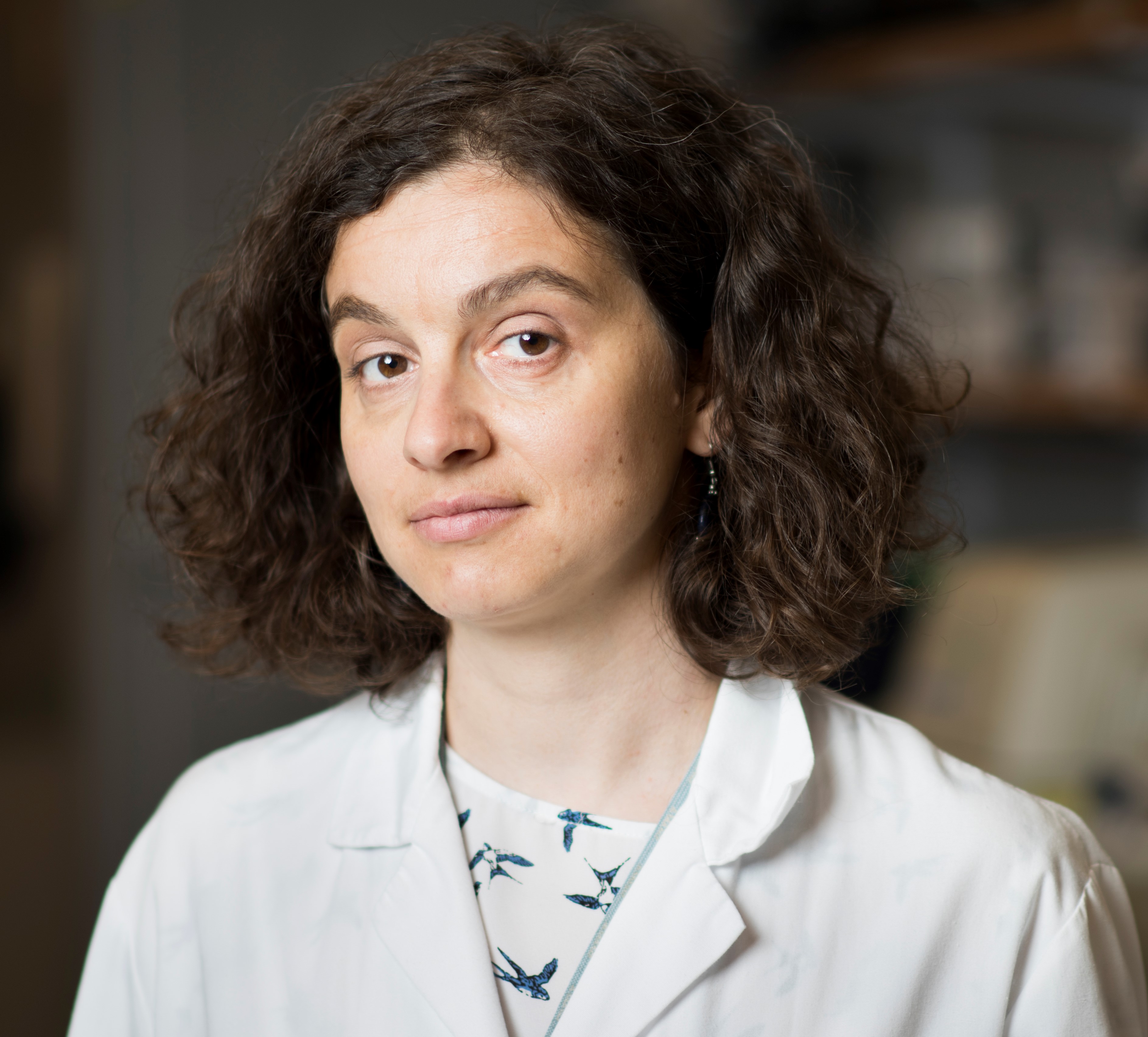
|
Marta Gaglia, Associate Professor
Tufts University, United States
Biography
Marta Gaglia is an associate professor of Molecular Biology and Microbiology at Tufts University School of Medicine. She received her PhD at University of California San Francisco. She developed her interest in virus-host interactions as a postdoctoral fellow at University of California Berkeley, where she began working on herpesviruses. She started her independent laboratory in 2014 at Tufts, where she continues to study how viruses repurpose host machinery for gene regulation and immune evasion in Kaposi’s sarcoma-associated herpesvirus and influenza A virus infections.
|
KSHV, Caspases and Immune Evasion: How KSHV Retools a Host Anti-Viral Pathway to Promote Its Replication
To replicate effectively, Kaposi’s sarcoma-associated herpesvirus (KSHV) strongly blocks induction of the type I interferon response in infected cells after reactivation of the lytic cycle. This inhibition of early anti-viral responses is likely to be crucial not only for viral spread and transmission but also for tumorigenesis, as viral replication contributes to the development of Kaposi’s sarcoma. While multiple viral proteins contribute to this block, our group has identified a surprising cellular factor with a key role in type I interferon inhibition, the cell death protease caspase-8. While caspases are generally considered anti-viral, caspase-8 appears to act in a pro-viral fashion and promote KSHV replication through inhibition of type I interferon induction. Our results suggest that caspase-8 is activated by the cellular Toll-like receptor pathway rather than through the canonical cell death signaling. Moreover, its activity does not lead to cell death, but instead inhibits the activity or activation of the cytoplasmic DNA sensor cGAS. Interestingly, using single-cell RNA sequencing we discovered that only a few cells express type I interferons during caspase inhibitor treatment, although the secreted interferon levels are high. These cells appear to be in the early lytic stage of reactivation and have higher level of expression of NF-kappaB proteins compared to the interferon-negative cells. Collectively, these findings indicate that KSHV hijacks one pathogen response pathway, the TLR pathway, to inhibit a different one, the viral DNA sensing pathway. KSHV also retools caspase-8, a cell death enzyme, to promote its own replication. In the absence of caspase signaling, a few reactivating cells turn on a strong type I interferon response that inhibits KSHV replication. We are now focusing on identifying the relevant targets of caspase-8 and deciphering what makes the interferon-producing cells unique.
|
|
| 8:45 - 9:00
|
|
Transition/Networking Break
|
| 9:00 - 10:30 - Concurrent Session
|
|
1A: Viral Entry and Egress I
Session Chairs:
Christine O'Connor, Assistant Professor
Case Western Reserve University , United States
|
Micah Luftig, Associate Professor and Co-Vice Chair
Duke University School of Medicine, United States
|
|
|
1B: Viral Replication
Session Chairs:
Gill Elliott, Professor
University of Surrey, United Kingdom
|
Rolf Renne, Professor
University of Florida, United States
|
|
|
1C: Virus-Host Interactions I
Session Chairs:
Ben Gewurz, Associate Professor
Brigham & Women's Hospital, Harvard Medical School, United States
|
Igor Jurak, Associate Professor
University of Rijeka, Croatia
|
|
| 10:30 - 11:00
|
|
Transition/Networking Break
|
| 11:00 - 11:40
|
|
Plenary 2 - Viral Genomics and Gene Expression
Session Chairs:
Neelam Sharma-Walia, Associate Professor
Rosalind Franklin University of Medicine and Science, United States
|
Robert F Kalejta, Professor, Oncology and Molecular Virology
University of Wisconsin-Madison, United States
|
Presenter:
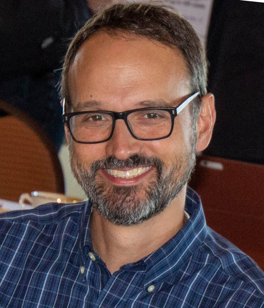
|
Joshua Munger, Associate Professor
University of Rochester Medical Center, United States
Biography
Joshua Munger is an Associate Professor in the Department of Biochemistry and Biophysics at the University of Rochester Medical Center. He obtained his Bachelor of Science degree at Virginia Tech and subsequently joined Bernard Roizman’s laboratory at the University of Chicago, where he obtained his doctoral degree. As a post-doctoral fellow, he worked in Thomas Shenk’s laboratory at Princeton University. Since joining the faculty at the University of Rochester his lab has studied the mechanisms through which Human Cytomegalovirus modulates cellular metabolism and alters innate immune signal transduction to support productive infection.
|
Human Cytomegalovirus Metabolic Remodeling: Harnessing Tissue Atypical Metabolic Isoforms to Support the Production of Infectious Virions
As obligate parasites, viruses depend on cellular metabolic resources to supply the energy and biomolecular building blocks necessary for their replication. Human Cytomegalovirus (HCMV) infection induces numerous metabolic activities that have been found to be important for productive infection. However, many of the mechanisms through which these metabolic activities are induced and how they contribute to infection are unclear. Through RNA-seq studies, we find that HCMV infection of fibroblasts induces several metabolic isoforms that are predominately expressed in other tissue types. Of these, the most substantially induced gene was the neuron-specific isoform of enolase (ENO2). Our results indicate that induction of ENO2 is important for HCMV-mediated glycolytic activation. Suppression of ENO2 induction attenuates the production of infectious virions. This defect appears very late in the viral life cycle and involves an enhanced production of non-infectious viral particles. Collectively, our data indicate that HCMV can harness a tissue atypical metabolic isoform to support metabolic remodeling and the production of infectious virions.
|
|
| 11:40 - 12:05
|
|
Symposium 2A
Presenter:

|
Michelle West, Professor
University of Sussex, United Kingdom
Biography
Professor Michelle West is Deputy Head of the School of Life Sciences (Research and Enterprise) at the University of Sussex, UK where she is a Professor of Tumour Virology. She obtained her Batchelors degree in Biochemistry from the University of Warwick and carried out her PhD work in the Department of Cancer Studies at the University of Birmingham working with Professor Martin Rowe on the regulation of transcription by Epstein-Barr virus (EBV). Professor West continued her gene regulation work at the University of Leicester working with Professor Anne Willis on translational regulation of the MYC oncogene. She returned to Virology in her second postdoctoral position at the Medical Research Council Laboratory of Molecular Biology in Cambridge working with Professor Jon Karn on transcriptional regulation by Human Immunodeficiency Virus 1. Professor West then established her own group at Sussex studying the molecular mechanisms involved in B cell transformation by EBV. Her research investigates how B cells are transcriptionally re-programmed by EBV with a focus on the structure and function of EBV transcription factors, long-range gene control and 3D chromatin interactions and cell cycle control mechanisms.
|
Three-dimensional Genome Rewiring by Epstein-Barr Virus
The human herpesvirus Epstein-Barr virus (EBV) is associated with the development of numerous B-cell lymphomas. Infection by EBV initially mimics short-term physiological B-cell activation but results in the outgrowth of latently-infected immortal B cells. EBV-encoded transcription factors (TFs) play crucial roles in B-cell immortalisation by epigenetically reprogramming the host cell genome to alter expression of large numbers of growth and survival genes. Gene deregulation by these viral TFs is predominantly mediated through the targeting of long-range regulatory elements. We have shown that at key lymphoma-associated genes EBV activators and repressors reconfigure enhancer-promoter interactions to activate or silence genes. To understand how EBV rewires enhancer-promoter interactions genome-wide to drive B-cell immortalisation and how this differs from events during short-term B cell activation, we have used high-throughput promoter capture (Capture Hi-C) in parallel with RNA-sequencing using EBV-infected CD40L/IL-4-activated B cells. Both EBV infection and CD40L/IL-4 B-cell activation were accompanied by key changes in 3D chromatin architecture: a reduction in long-range promoter interactions and localised decondensation of chromatin. For differentially expressed genes, we found that the number of promoter-long range interactions correlated with the change in gene expression linking gene regulation to promoter contact changes. We also identified novel interaction hubs comprising EBV-specific or CD40L/IL-4 specific co-regulated gene clusters. Our data provide new insights into 3D promoter rewiring during B-cell activation.
|
|
| 12:05 - 12:30
|
|
Symposium 2B
Presenter:

|
Jill Dembowski, Assistant Professor
Duquesne University, United States
Biography
Dr. Dembowski is an Assistant Professor in the Department of Biological Sciences at Duquesne University. She received her Ph.D. in Molecular and Cell Biology from the University of Pittsburgh for her work on the investigation of alternative pre-mRNA splicing regulation in the nervous system. She then performed postdoctoral work at Carnegie Mellon University where she studied mechanisms of eukaryotic ribosome assembly. She then joined the laboratory of Dr. Neal DeLuca at the University of Pittsburgh School of Medicine where she was promoted to Research Assistant Professor. There she developed approaches to purify herpes simplex virus genomes from infected cells for the identification of associated proteins by mass spectrometry. These studies revealed the dynamics of viral and host protein interactions with HSV-1 genomes throughout the course of lytic infection, as well as host proteins that selectively associate with viral replication forks. Now at Duquesne University, the Dembowski lab focuses on investigating the roles viral genome associated cellular proteins play in viral DNA replication, repair, and gene expression.
|
New insights into the roles of host factors in HSV-1 DNA replication, genome maintenance, and late gene expression
Much of the HSV-1 life cycle occurs in the nucleus of infected cells, where events that occur on the viral genome determine the outcome of infection. HSV-1 exploits host factors for viral gene expression and activates host DNA damage responses, while viral factors inhibit downstream steps in DNA damage response pathways. Herpesviral DNA replication is coupled to transcription, DNA recombination, and DNA repair, although the mechanisms by which these processes are coordinated and regulated on the DNA are not understood. The recent development of proteomic approaches to study protein association with replication forks has enabled an in depth look at this fundamental process within cells. In a previous study, we selectively purified HSV-1 replication forks and associated proteins from virus-infected cells. In addition to the viral replication machinery, we found that select host chromatin remodeling, transcription, DNA modifying, and DNA repair factors associate with HSV-1 replication forks. Our long-term goal is to determine how these host factors are selectively recruited to viral replication forks and what roles they play in promoting or inhibiting viral replication or coupled processes. To this end, we have demonstrated that inhibition of cellular factors found at or near viral replication forks including the cellular processivity factor proliferating cell nuclear antigen (PCNA) and lysine specific demethylase LSD1 results in selective inhibition of HSV-1 DNA replication and subsequent late gene expression. In ongoing experiments, we are investigating how viral replication is stalled in the absence of PCNA and LSD1. We anticipate that these studies will provide insight into mechanisms that couple HSV-1 DNA replication with DNA repair and transcription for the coordinated maintenance and expression of the viral genome.
|
|
| 12:30 - 12:45
|
|
Transition/Networking Break
|
| 12:45 - 13:25
|
|
Priscilla Schaffer Lecture
Session Chair:
Angus Wilson, Associate Professor
NYU Grossman School of Medicine, United States
|
Presenter:

|
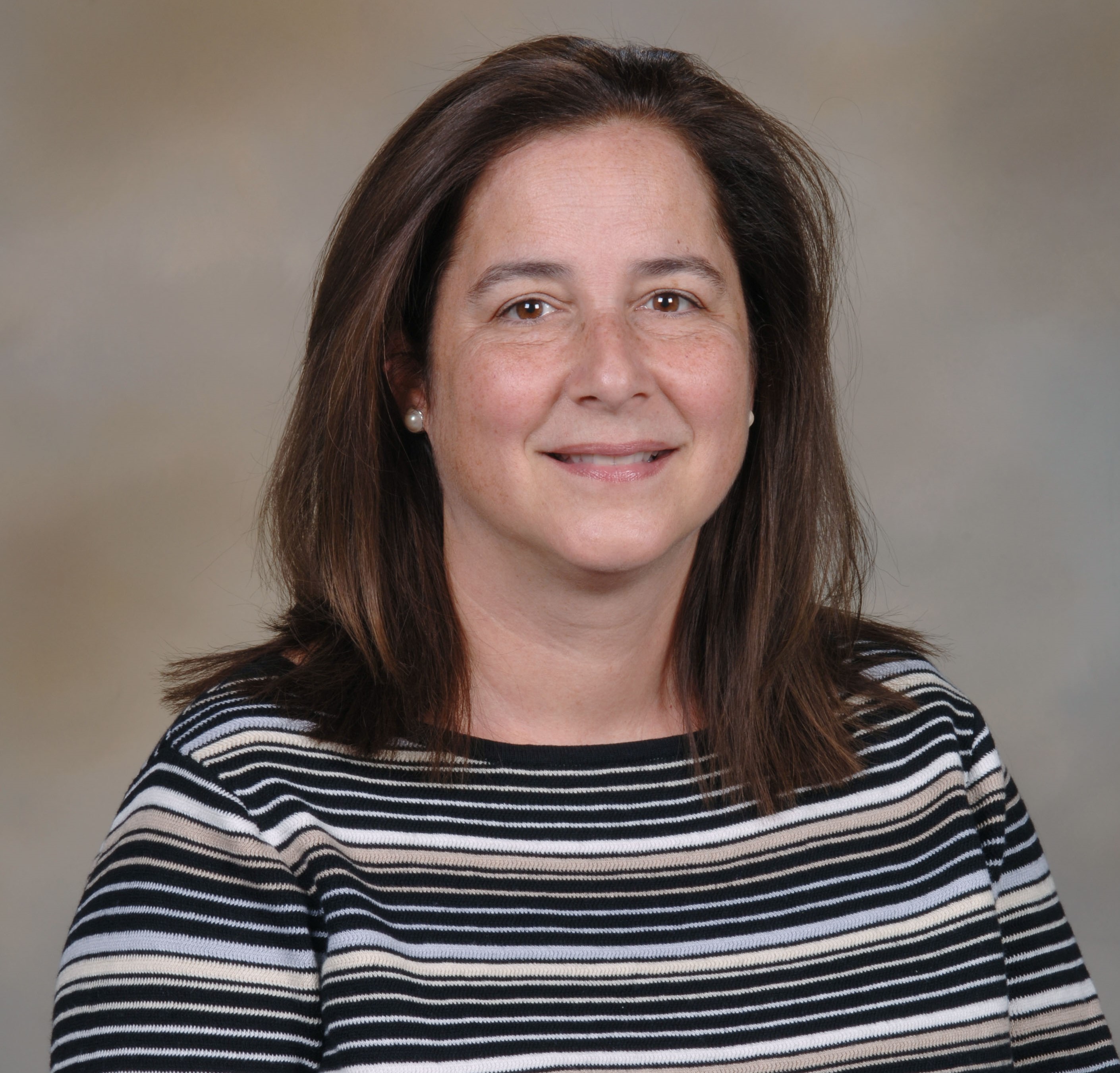
Anna Cliffe, Assistant Professor
University of Virginia, United States
Biography
Anna Cliffe is an Assistant Professor of Microbiology, Immunology and Cancer Biology at the University of Virginia (UVa) in Charlottesville, Virginia. She received her PhD from the University of Edinburgh, where she investigated the role of viral tRNA-like molecules in MHV-68 infection. Upon completing her PhD, she moved to Dr. David Knipe's laboratory at Harvard Medical School to investigate the chromatin control of HSV-1 lytic replication and latency. She then moved to Dr. Mohanish Deshmukh's laboratory in the Neuroscience Center at UNC Chapel Hill, where she used a primary neuronal model of HSV-1 latency to investigate how neuronal stress triggers reactivation. Using multiple model systems including primary neuronal cultures, induced human neurons and animal models, research in her lab aims to understand how viral gene silencing occurs in neurons, the intersection between the viral genome and host immune signaling and the contribution of HSV infection to the development of neurodegenerative disease.
|
Intersection of the Herpes Simplex Virus Genome with Neuronal Innate Immune Signaling Pathways
Herpes simplex virus (HSV) establishes a latent infection in neurons, where it persists for life and periodically reactivates to cause disease. During latency, the viral episome assembles into silent heterochromatin, which is remodeled for reactivation to occur. The viral genome can be regulated epigenetically during the establishment, maintenance and reactivation from latency. We aim to understand how the nature of gene silencing is modulated in neurons to result in different forms of silencing and how silencing is reversed for reactivation. Previously, we identified a role for a neural stress pathway involving DLK-mediated JNK-activity in HSV reactivation triggered by nerve growth factor (NGF) deprivation. Reactivation was associated with a JNK-dependent histone phospho/methyl switch on lytic gene promoters. Given that the same histone phospho/methyl switch occurs in cortical neurons following hyperexcitability, we examined whether HSV reactivation was linked to hyperexcitability and the contribution of JNK activity and histone phosphorylation. We found that reactivation was induced by neuronal hyperexcitation. Moreover, reactivation was dependent on DLK/JNK activity and linked to a histone phospho/methyl switch. We next investigated whether physiological triggers induce HSV reactivation via hyperexcitability. Notably, IL-1 induced sympathetic neurons to enter a hyperexcitable state. IL-1 also triggered DLK/JNK-dependent HSV reactivation that was dependent on neuronal activity. Therefore, HSV has highjacked an innate immune pathway to induce reactivation. These data have relevance for the potential mechanism of reactivation in response to psychological stress, fever and UV damage, which are all linked with IL-1 release. We have also established that IL-1 signaling is not the only innate immune pathway used by HSV for reactivation, as data indicate that HSV also co-opts the nucleic acid sensing pathways during reactivation. The co-option of IL-1 signaling and nucleotide sensing pathways in reactivation are, however, in contrast to the role of other anti-viral mechanisms that promote latency and/or restrict reactivation. Notably, treatment of neurons at the time of infection with type I or type II IFN results deep forms of latency that are restricted for reactivation. For type I IFN, we have found that this results in prolonged association of viral genomes in PML nuclear bodies. In addition, reactivation could be rescued by PML depletion. Thus, the viral genome has a memory of the conditions upon de novo infection to result in a more restricted form of latency. This work highlights how specific innate immune signaling pathways in neurons converge on the viral genome to differentially regulate HSV latency and reactivation.
|
|



.jpg)
